
Our paper on the first region-of-interest high resolution X-ray diffraction computed tomography experiment of a Si-graphite electrode used for Li-ion battery applications has been published in Nano Letters
Our scientists’ new work on Si-graphite electrodes used for Li-ion battery applications with high resolution in situ X-ray chemical imaging has been published in a new paper, “Spatially Resolving Lithiation in Silicon–Graphite Composite Electrodes via in Situ High-Energy X-ray Diffraction Computed Tomography” in Nano Letters. The work was performed with Donal Finegan from the National Renewable Energy Laboratory and in collaboration with a team from the Electrochemical Innovation Lab (EIL) from UCL Chemical Engineering using ESRF’s ID15A beamline.
Optimizing the chemical and morphological parameters of lithium-ion (Li-ion) electrodes is extremely challenging, due in part to the absence of techniques to construct spatial and temporal descriptions of chemical and morphological heterogeneities. In this work, we present the first demonstration of combined high-speed X-ray diffraction (XRD) and XRD computed tomography (XRD-CT) to probe, in 3D, crystallographic heterogeneities within Li-ion electrodes with a spatial resolution of 1 μm. The local charge-transfer mechanism within and between individual particles was investigated in a silicon(Si)−graphite composite electrode. High-speed XRD revealed charge balancing kinetics between the graphite and Si during the minutes following the transition from operation to open circuit. Subparticle lithiation heterogeneities in both Si and graphite were observed using XRD-CT, where the core and shell structures were segmented, and their respective diffraction patterns were characterized.
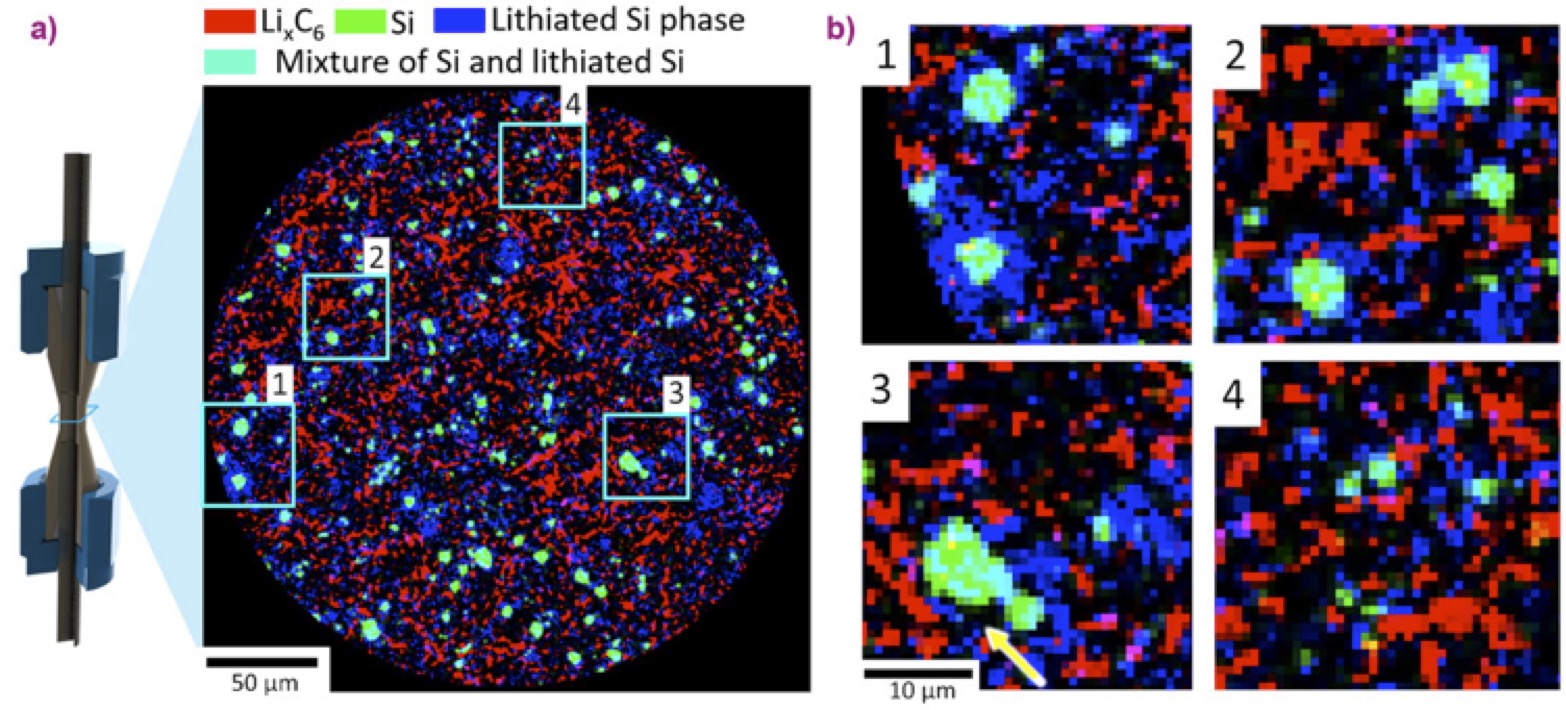
Figure: (a) XRD-CT slice taken at the beginning of the charge step showing a phase-distribution map of LiC12 (red), crystalline Si (green), and lithium silicides LixSi (blue). According to additive colour mixing, the colour teal represents a mixture of green (Si) and blue (lithiated Si). (b) Magnified regions of interest showing large particles of LixSi phase with crystalline Si cores (1-3) and smaller LixSi particles (4) interspersed in the graphite matrix. The yellow arrow highlights what looks to be evidence of delamination from a crystalline Si core.
Read more about it at https://www.esrf.eu/home/news/spotlight/content-news/spotlight/spotlight346.html
Read the article at https://pubs.acs.org/doi/full/10.1021/acs.nanolett.9b00955